What is an MRI?
Magnetic resonance imaging (MRI) is a diagnostic test that uses a powerful magnet and radio wave pulses to create detailed images of body tissue. The test does not require entering the body (it is noninvasive). The images produced by MRI allow doctors to clearly see changes in brain tissue and blood flow that are too small to see on a CT scan or ultrasound. MRI tests are used to identify damage caused to the brain by stroke, including bleeding and blood flow problems caused by blood clots or blood vessel abnormalities. An MRI can also rule out other diseases of the brain, such as tumors.
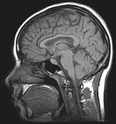
An MRI image of the head
What is MRA?
Magnetic resonance angiography (MRA) is a special type of MRI test that focuses on blood flow and the blood vessels of the brain, instead of on all brain tissue like a standard MRI. It can detect narrowing or blockage of the blood vessels inside and outside the brain—such as the carotid arteries in the neck—especially when it is used together with carotid or transcranial Doppler ultrasound. MRA can also be used to confirm a blockage seen in other MRI scans. MRA is useful in detecting weakening or ballooning of the blood vessels (aneurysms), which can lead to bleeding in the brain.1 Some, but not all, MRA tests use injections of a special dye to produce more detailed images of the blood vessels in the brain.
What other types of MRI tests are there?
The two main variations of a standard MRI are diffusion MRI and perfusion MRI. These tests use an MR imaging machine and special techniques to process the data differently, highlighting specific features of the brain tissue.
Diffusion MRI measures the movement of water in the brain. Under normal conditions, water moves freely; when blood supply to the brain is interrupted, brain tissue swells and slows the movement of water. A diffusion MRI scan can pinpoint which area of the brain is getting little or no blood supply within minutes of a stroke, faster than any other imaging test.2 It can also measure how much brain tissue was damaged by the stroke and how recently the damage occurred. When diffusion MRI is available at the hospital it may be used as the first test to diagnose acute stroke.2
Perfusion MRI uses a rapid injection of a small amount of dye that circulates in the bloodstream and shows if your brain is receiving enough blood. It can generate a map of the amount and speed of blood flowing throughout the brain. Perfusion MRI can be done together with diffusion MRI to provide information about how much brain tissue has been damaged permanently and how much can still be saved.3 However, the perfusion MRI maps take extra time, and for now it is an optional test to evaluate an acute stroke.2
Who might have a MRI or MRA test?
A CT scan is the standard initial test to find out if a stroke was caused by bleeding or a blocked vessel, mainly because it is available at all hospitals and is fast. An MRI scan is better than CT at detecting the brain changes caused by a stroke, but the MRI test takes slightly longer to perform, about 30 to 45 minutes (compared to 20 minutes for a CT scan). In hospitals where it is available, MRI is an effective first test for diagnosing a stroke and identifying bleeding in the brain, as long it does not delay treatment too much.4 Some hospitals have stroke-specific MRI tests that take 15 to 20 minutes.3 After the initial diagnosis has been made, an MRI scan is useful in determining what caused the stroke and how much brain tissue can be saved, helping your doctor to make an accurate prognosis and determine the best treatment for you.
MRA can detect blood vessel abnormalities or problems causing reduced or blocked blood flow to the brain and putting you at risk for a first or second stroke. If a carotid Doppler ultrasound has found narrowing or blockage in the arteries on each side of your neck, an MRA may be used to confirm the results. An MRA is a more sensitive test to help determine if you would benefit from treatments such as carotid endarterectomy and carotid stenting to reduce your risk for TIA or stroke.
If you have polycystic kidney disease or a family history of brain aneurysms and bleeding in the space around the brain (subarachnoid hemorrhage), your doctor may order an MRA to see if you have an aneurysm so steps can be taken to correct it before it causes a stroke. One study found that people with two or more first-degree relatives (parents, children, or siblings) with subarachnoid hemorrhage may benefit from aneurysm screening.5 The accuracy of the MRA depends on the size of the aneurysm: smaller ones are harder to detect. People who are not at high risk for aneurysms should not be routinely screened because the risks of the test may outweigh the benefits.


