There are three situations in which tests are used to diagnose stroke and guide your medical care:
- Diagnosing a stroke and telling the two main types of stroke apart
- Finding out what caused the stroke once it has been diagnosed
- Evaluating your risk for a first or repeat stroke
Some tests are used in more than one situation. Click on the name of any test to go directly to the complete article for more information.
If you suspect you have had a stroke, the doctor will first ask you about your medical history and your stroke symptoms and perform a physical exam and a neurological exam to determine how your body, mind, and nervous system are working.
The main priority for patients with a suspected stroke is quickly determining if you have had a stroke, and, if so, whether it is a blocked-vessel (ischemic or embolic) stroke or a bleeding (hemorrhagic) stroke. The treatments for these two types of stroke are very different. The sooner doctors know you have a stroke and what type it is, the sooner you can be treated.
The most common test to tell the two main types of stroke apart is the computerized tomography (CT) scan (sometimes also called a CAT scan). A CT scan can also tell if something else might be causing the stroke symptoms, such as a brain tumor. Some hospitals use a magnetic resonance imaging (MRI) scan as the initial imaging test.
You will also have blood tests to provide general information about your health and to rule out other causes of stroke.
Finding Out What Caused the Stroke
Once a stroke is diagnosed, it is important to find out where exactly the stroke is in the brain and what caused it. This allows doctors to determine the best treatment and how to help you prevent future strokes. Note: if you have had a blocked-vessel stroke and are eligible for first-line treatment with the clot-busting tissue plasminogen activator (tPA), you should receive it before any more tests are performed.
Tests that look at your heart, such as an ECG or an echocardiogram, may be performed to find out if you have heart problems such as atrial fibrillation or valve disease that could have caused a blood clot to form in the heart. Clots that form in the heart can travel to the brain, causing an embolic stroke. Blood tests can also help doctors rule out cardiac and other possible causes of stroke.
Other tests can determine the location and size of any blockages in the vessels that supply blood to your brain. A carotid Doppler ultrasound looks at the arteries of the neck (the carotid arteries) to see if you can benefit from treatment to open these vessels and prevent another stroke. A transcranial Doppler ultrasound can measure blood flow in the large arteries in your brain. Tests that look at the large and small blood vessels in your brain include angiography of the head and neck, magnetic resonance angiography (MRA), and advanced versions of the CT scan such as CT angiography and perfusion CT. These tests can determine if you have blockages in the arteries in your brain and can also detect any other problems that could have caused the stroke, such as an aneurysm.
If you have had seizures or you doctors suspects you have, your brain’s electrical activity will be monitored with an EEG (electroencephalogram).
Figure 1: Imaging Tests In Stroke Diagnosis and Treatment

Evaluating Your Risk for Stroke
If you have already had a stroke or transient ischemic attack (TIA), you are automatically at high risk for another stroke and should take steps to prevent it. See “Steps to Prevent Another Stroke” in our Recovery section for more.
If you have never had a stroke or TIA, use our stroke risk calculator for a rough estimate of your risk for stroke. Tools that help doctors evaluate and control your stroke risk include testing for stroke risk factors, such as blood pressure and cholesterol measurements, a glucose test to diagnose diabetes, and other blood tests to measure the overall health of your blood.
The C-reactive protein test and the PLAC test detect proteins that contribute to the inflammatory process of atherosclerosis. These tests can help clarify your stroke risk and determine how aggressively you should be treated if you are between high and low risk for stroke.
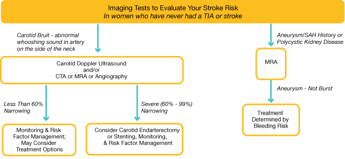
Patients at high risk for stroke may have a carotid Doppler ultrasound to see if narrowing or blockages in the arteries in the neck are putting you at risk for stroke. If they are, treatment or surgery can help prevent a future stroke. Patients who have a family history of strokes caused by aneurysms may have an MRA or an angiogram to identify any developing problems before they cause a stroke.
Figure 2: Imaging Tests to Evaluate Your Stroke Risk

Click here to continue on to our first stroke testing and diagnosis article, the Physical and Neurological Exam.


