What is angiography?
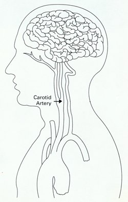
Angiography is a diagnostic test that produces an X-ray movie of the blood vessels and the blood flowing through them. Angiography of the head (cerebral angiography) is used to diagnose stroke by revealing abnormalities such as narrowing or blockage of the brain’s blood vessels or bleeding in the brain. Angiography of the neck (carotid angiography) looks at the two large carotid arteries—one on each side of the neck—that supply blood to the brain. Narrowing in the carotid arteries reduces blood flow, and fatty deposits on the artery walls can break off, causing a stroke.

The location of the carotid artery on one side of the neck (NINDS)
To perform the test, a long, thin tube called a catheter is inserted into an artery in your groin or arm and guided to the area to be studied. The catheter is used to inject a dye into the blood vessels of your head and neck, and the dye is filmed by an X-ray camera, producing an angiogram—an X-ray movie of your vessels as the blood moves through them.

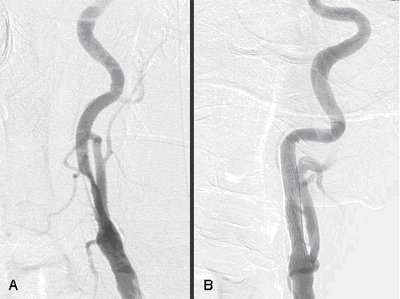
Angiogram image of a narrowed carotid artery (A) and the same artery after it was opened with a stent (B)
Are there any alternatives to invasive angiography?
This section refers to a conventional angiogram, in which a catheter is used to inject dye into the vessels. There are less invasive alternatives that do not require a catheter or an incision, such as CT angiography or Magnetic Resonance Angiography (MRA). These tests can also be used to take a close look at the cause of a stroke and decide treatment options in patients who cannot or chose not to undergo conventional angiography. The pictures produced by these tests, although detailed and accurate enough for most uses, are not quite as precise as the ones from a conventional angiogram.
Who might have a head or neck angiogram?
Angiography is the gold standard for detecting problems with the blood vessels of the head and neck because it produces pictures with a degree of detail not available from other tests.1 Because it requires entering the body (is invasive) and involves some risks, head angiography is usually done when a previous noninvasive test, such as a CT scan or MRI or ultrasound, has detected an abnormality and more information is needed.2
Carotid (neck) angiography is used to look for a blockage or narrowing of the carotid artery to decide if you would benefit from a procedure to prevent stroke, such as a carotid endarterectomy or carotid stenting.
Cerebral (head) angiography is often performed if you have had a TIA or a stroke and doctors want to determine if treatment is required, or to see if treatment has been effective at opening a blocked vessel.


